Gang Zhao1,
Xiao-Hui Wang1 ![]() ,
Ren-Qian Song1,
Chun-Lin Zhang2
,
Ren-Qian Song1,
Chun-Lin Zhang2
For correspondence:- Xiao-Hui Wang Email: wangxiaohuiw@hotmail.com Tel:+86317666666
Received: 1 July 2015 Accepted: 6 February 2016 Published: 31 March 2016
Citation:
Zhao G, Wang X, Song R, Zhang C.
Inhibition of caspase-3, -6, -8 and -9 ex
© 2016 The authors.
This is an Open Access article that uses a funding model which does not charge readers or their institutions for access and distributed under the terms of the Creative Commons Attribution License (http://creativecommons.org/licenses/by/4.0) and the Budapest Open Access Initiative (http://www.budapestopenaccessinitiative.org/read), which permit unrestricted use, distribution, and reproduction in any medium, provided the original work is properly credited..
Purpose: To demonstrate the anti-apoptotic effects of cantharidin in mice with acute spinal cord injury (ASCI).
Methods: In total, 30 male Sprague-Dawley mice were divided into three groups of 10 animals each. ASCI was induced in two of the groups using a modified Allen's method, consisting of treatment with 10 mg/kg body weight cantharidin after injury, and sacrifice on days 2, 5, 10, 20 and 30 to extract the spinal cord. The activity levels of caspase-3, -6, -8, and -9 were determined spectrophotometrically at 455 nm with a microplate reader.
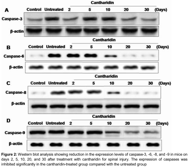
Results: The results showed that cantharidin treatment caused a significant reduction in the ex
Conclusion: Cantharidin treatment exerts an anti-apoptotic effect against secondary spinal cord injury (SCI) by suppressing caspase activity. Thus, cantharidin may be suitable for the treatment of secondary SCI.
Introduction
Spinal cord injury (SCI) characterised by a break in the spinal cord is among the most severe health problems worldwide and is caused mainly by traffic accidents [1,2]. Two phases of SCI have been defined: the acute phase, resulting from compression and haemorrhage of tissues, and the secondary phase, which is accompanied by cell apoptosis at the lesion [1,2]. The secondary injury phase is usually more problematic and severe than the primary injury phase, leading to long-lasting disability. Because nervous system tissues do not possess the ability to regenerate, treatment should be free from side effects, and should be administered systemically [3]. Early treatment of acute spinal cord injury (ACSI) involves protection of the uninjured spinal cord, prevention of secondary injury, and promotion of spinal cord function. Many molecules have been used to inhibit the process of cell apoptosis in neurological disorders, such as traumatic brain injury [3,4]. The inhibition of apoptosis in the spinal cord has been reported to play an important role after ASCI [5]. For the inhibition of apoptosis, suppression of caspase activation by chemotherapeutic agents can be highly effective.
Cantharidin, a terpenoid isolated from the blister beetle, a member of the Meloidae family and Coleoptera order [6], has been used in traditional Chinese medicine for the treatment of various disorders [7]. Cantharidin treatment had been shown to induce cell cycle arrest in cancer cells [8]. In addition, cantharidin treatment induces apoptosis in different types of cancer cell, including hepatoma [9], colon [10], bladder [11], breast [12], and leukaemia [13] cells. Furthermore, in T24 cells, treatment with cantharidin has been shown to induce over-expression of COX2 and production of PGE2 [11].
In the present study, the effect of cantharidin on the spinal cord after ASCI was studied in mice. The results demonstrated that cantharidin treatment inhibited the expression of caspase-3, -6, -8, and -9. These caspases are known to play an important role in the process of cell apoptosis.
Methods
Chemicals and reagents
Cantharidin and dimethyl sulphoxide were obtained from Sigma Chemical Co. (St. Louis, MO, USA). Penicillin injections were purchased from Gibco BRL (Grand Island, NY, USA).
Animals
Adult male Sprague-Dawley mice (~190–210 g) were obtained from the Shanghai Laboratory Animal Commission (Shanghai, China). All experimental procedures were performed according to the Guidance for the Care and Use of Laboratory Animals (2012) of the Ministry of Science and Technology of the People's Republic of China, and ethical approval was granted by the institutional ethical committee. The animals were acclimatised to the laboratory conditions 1 week before the experiment was started and were housed in a room with 12-h dark/light cycle.
Preparation of the ASCI animal model and treatment strategy
In total, 30 animals were divided randomly into three groups of 10 animals each: (a) a control group without SCI, (b) a group of animals with SCI that received no treatment, and a group of animals with SCI treated with cantharidin. Cantharidin was administered to each animal at a concentration of 10 mg/kg body weight after injury. Animals in control and untreated groups received an equal volume of normal saline.
Sample collection
The animals were sacrificed on days 2, 5, 10, 20, and 30 after injury to extract the spinal cord. For the purpose of spinal cord extraction, a small incision was made on the dorsal side after the animal was anaesthetised, followed by removal of skin and other subcutaneous tissues. After exposure, the spinal cord was removed carefully and placed in lysis buffer (40 mmol/l Tris HCl, 1 mmol/l EDTA, 150 mmol/l KCl, 100 mmol/L NaVO3, 1 % Triton X 100, and 1 mmol/L PMSF; pH 7.5) for homogenation.
The homogenised tissues were put into tubes (1.5 mL) and centrifuged for 45 min at 12,000 rpm. Following centrifugation, the solid material was removed and put into the centrifuge tube. The tubes were then stored at –80 °C for determination of caspase levels.
ELISA for determination of caspase activity
An enzyme-linked immunosorbent assay (ELISA) kit (DVE00; R&D Systems, Minneapolis, MN, USA) was used for the determination of caspase activity according to the manufacturer’s protocol. Briefly, dilution solutions were prepared with 1.8 mL detection buffer and 0.2 mL lysis buffer. Dilution combinations – 0, 10, 20, 50, 100, and 200 μM 4-nitroaniline – were prepared and used as controls.
Absorbance at 455 nm was recorded for 100-μL samples. The absorbance values for blank wells were subtracted from those of the standard samples (Chemicon, Temecula, CA, USA) with respect to caspase-3 activity. Similarly, absorbance was recorded for the caspases.
Western blotting for apoptosis-associated proteins
The spinal cord tissues were lysed in lysis buffer containing 40 mM Tris-HCl (pH 7.4), 10 mM EDTA, 120 mM NaCl, 1 mM dithiothreitol, and 0.1 % Nonide P-40. Total proteins were separated on 10 % Tris-glycine SDS-polyacrylamide gels and then transferred to a nitrocellulose membrane by electroblotting. The membranes were incubated with primary antibodies for caspase-3, -6, -8, and 9 (R&D Systems).
After incubation, the membranes were washed with PBS, followed by incubation with secondary antibodies for enhanced chemiluminescence (NEN Life Science Products, Inc., Boston, MA, USA). The anti-ß-tubulin from a mouse monoclonal antibody was used as the loading control.
Statistical analysis
SPSS for Windows software package (ver. 18.0; SPSS Inc., Chicago, IL, USA) was used for statistical analysis of the data obtained. The data are presented as mean ± standard error of the mean (SEM). For comparison of means among groups, one way analysis of variance and post-hoc least significant difference test were used. Differences were considered statistically significant at p < 0.05.
Results
Using ELISA and western blot analyses in the control group, the activity of caspase-3, -6, and -8 was determined to remain similar throughout the treatment period.
Examination of the expression of caspase-3, -6, -8, and -9, using ELISA and western blot analyses in the treatment group revealed that administration of cantharidin caused a marked reduction in the expression of caspases compared with the untreated rats on days 2, 5, 10, and 20. However, on day 30, the expression levels of caspases in the treated and untreated ACSI groups were similar and close to those of the control group (Tables 1 – 4, ).
Discussion
Cantharidin, which has been used in traditional Chinese medicine, induces arrest of the cell cycle in cancer cells [7,8]. In addition, cantharidin treatment of different types of cancer cell, including hepatoma [9], colon [10], bladder [11], breast [12], oral buccal and leukaemia [13] cells, has been shown to induce apoptosis. The present study was performed to investigate the effect of cantharidin on apoptosis in the spinal cord after injury in rats. The results demonstrated that cantharidin treatment inhibited the expression of caspase-3, -6, -8, and -9, which together represent the vital proteases involved in the process of cell apoptosis.
Cellular apoptosis plays an important role in the human body by removing unwanted cells; however, in patients with SCI, it leads to the development of secondary injury. Apoptosis is initiated and regulated by various factors, including caspases, Bcl 2, and Bax [14–16]. Studies have shown that increased expression of caspases results in the induction of apoptosis [17]. Among the various known caspases, initiator caspases (caspase-2, -8, -9, and -10) begin the apoptotic process. Caspase 3 has also been reported to be the most important initiator caspase, and to play a vital role in initiating apoptosis. The expression of caspase-3 induces various proteins, such as PAK2, mdm2, D4-GDI, and PARP, which in turn induce cell apoptosis. The data from the present study demonstrate that the expression of initiator (caspase-8 and -9) and effector (caspase-3 and -6) caspases was reduced in rats treated with cantharidin on days 2, 5, 10, and 20.
Previous studies have shown that caspase-3 activation in turn activates caspase-8 through a sequence of changes [18]. However, the results from our study revealed that cantharidin treatment inhibited the expression of caspase-8, which may have been due to a reduction in the expression of caspase-6.
Caspase-9, another member of the initiator caspase family, is involved in inducing apoptosis through the mitochondria. Our results showed that administration of cantharidin also reduced the expression of caspase-9; these results were verified by western blot analysis. These findings support the ability of cantharidin treatment to prevent the development of secondary spinal injury in mice through the inhibition of cell apoptosis via a mitochondrial pathway.
Conclusion
Cantharidin treatment inhibits secondary injury in the spinal cord by suppressing caspase expression, which can occur via a mitochondria dependent apoptotic pathway. Therefore, cantharidin can be used for the treatment of secondary SCI.
References
Archives


News Updates